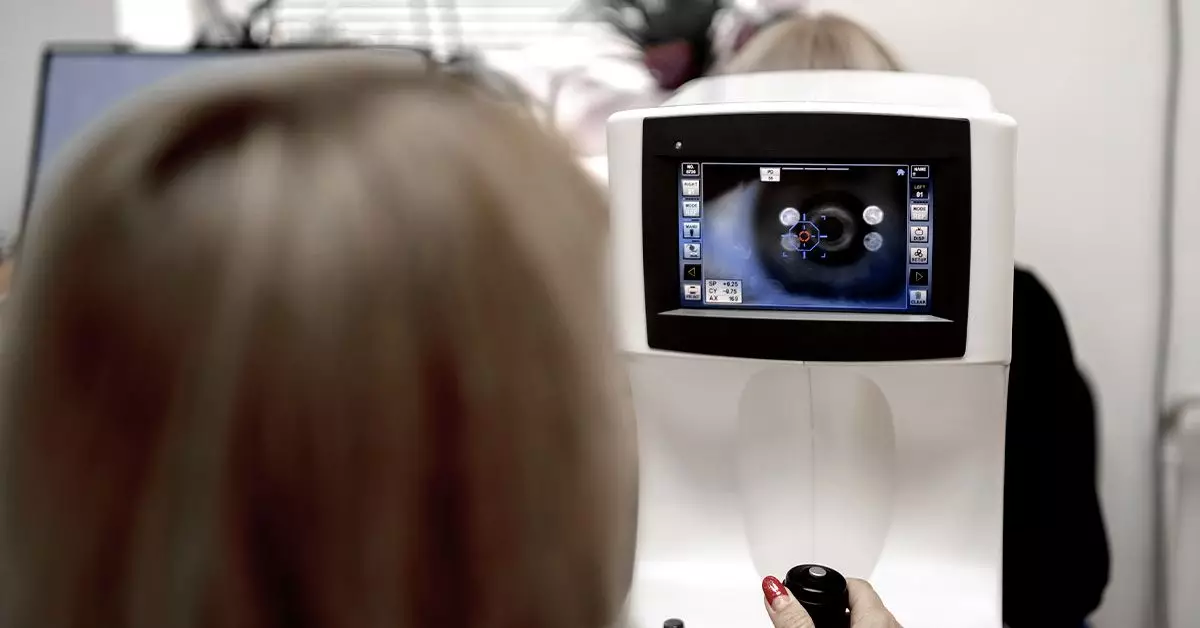
The preservation of vision is a critical aspect of healthcare, especially for those living with diabetes. Among the various methods employed for eye health assessments, fundus photography has emerged as a vital technique for diagnosing conditions like diabetic retinopathy. This article delves into the significance of fundus photography, the process of capturing these crucial images, and the implications for patients dealing with diabetes.
Fundus photography involves capturing images of the inner structures of the eye, particularly the retina. This imaging technique is a subset of fundoscopy, which refers to the comprehensive examination of the fundus (the interior surface at the back of the eye). The primary objective of fundus photography is to identify any pathological changes that may indicate the presence of eye diseases such as diabetic retinopathy, age-related macular degeneration, or glaucoma.
The process of fundus photography is straightforward: an eye care professional uses a specialized camera to photograph the retina. This photographic record is invaluable not only for diagnosis but also for monitoring disease progression and the effectiveness of treatments over time. It provides a detailed view of specific eye conditions that may otherwise go unnoticed without intervention.
Diabetic retinopathy is a significant complication arising from diabetes, with the potential to lead to severe vision impairment or blindness if left untreated. The early stages of diabetic retinopathy often do not manifest noticeable symptoms, making regular screenings critical. Fundus photography plays an indispensable role in identifying the telltale signs of this condition.
When analyzing fundus images for diabetic retinopathy, eye care professionals look for key indicators such as microaneurysms, which appear as small red dots, indicating the initial stages of vascular damage. Other indicators include retinal hemorrhages—either dot-and-blot or flame-shaped—which shed light on bleeding within the retina due to weakened blood vessels. Moreover, hard exudates, cotton wool spots, and signs of neovascularization signify the advanced progression of the disease, leading to potentially irreversible vision loss if not addressed.
To successfully perform fundus photography, the pupils need to be dilated. This procedure typically involves the use of eye drops that relax the muscles controlling pupil size. The dilation process can take approximately 20 to 30 minutes, and after the exam, some individuals may experience temporary blurred vision and increased sensitivity to light. Patients are generally advised not to drive immediately following the test, as their vision may remain affected for several hours.
Despite its simplicity and efficiency, the process is not without its drawbacks. The use of eye drops can lead to discomfort, and individuals may experience side effects. Therefore, it is important for patients to understand what to expect and to plan accordingly.
Advances in technology have led to innovative methods of conducting fundus photography, notably through smartphone technology. Selfie Fundus Imaging (SFI) allows individuals to capture fundus images using their smartphones. Preliminary studies suggest that SFI can yield results comparable to traditional methods performed by trained technicians.
This development has significant implications for accessibility and cost-effectiveness in screening for diabetic retinopathy. By enabling patients to take their own fundus photos, SFI may help overcome barriers related to distance from specialized services and reduce the burden on healthcare systems.
For individuals with diabetes, the risk of developing diabetic retinopathy underscores the importance of regular eye examinations. Eye care professionals recommend that patients with diabetes undergo dilated eye exams at least once annually to monitor for changes. Pregnant women with diabetes or those with gestational diabetes should seek immediate eye evaluations, given the increased risk during this time.
Earlier detection and management of diabetic retinopathy can significantly lower the risk of vision loss. Research indicates that timely intervention can prevent approximately 90% of blindness associated with diabetic retinopathy. Consequently, maintaining a balanced lifestyle that includes regular exercise, a nutritious diet, and strict adherence to medical regimens is critical in mitigating complications.
Fundus photography stands as a vital component in the early detection and management of diabetic retinopathy, ultimately preserving vision and improving the quality of life for those affected by diabetes. Regular eye examinations and advancements such as Selfie Fundus Imaging provide accessible pathways to ensure ongoing monitoring of eye health. By understanding the significance of these screenings and taking proactive steps, individuals can safeguard their vision against the deleterious effects of diabetic retinopathy.


Leave a Reply